Included Teeth
What are included teeth?
Sometimes the teeth fail to erupt in the mouth, once they are formed inside the jaws, being included within them. They are the so-called included teeth. Depending on the depth to which they are buried, we speak of total or partial inclusions. If they are partial inclusions, there may be a communication of the tooth with the mouth, saliva and bacteria present in them and be the cause of recurrent infections.
Tooth inclusions are often accompanied by malposition of the tooth, so that they can be impacted against neighboring teeth. This impaction can cause irreparable damage to neighboring teeth such as:
- Root reabsorption (rhizolysis)
- Bone resorption around the root, creating periodontal pockets
- Caries formation and development of cysts and tumors in relation to the tissues that originally formed the included tooth


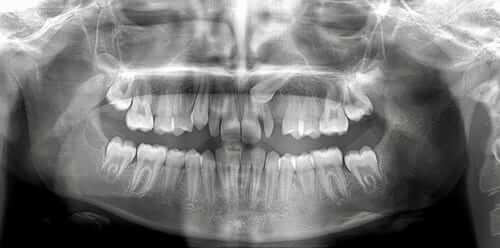
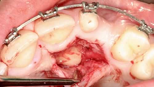
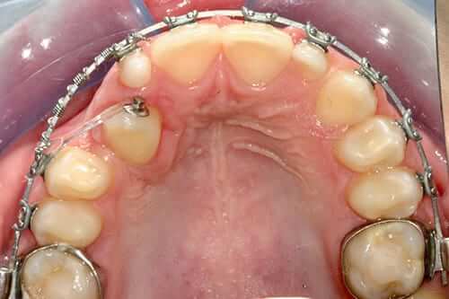
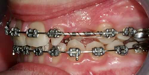
We present a case with a canine totally included in the palate and with an inclination that made it physiologically impossible to exit the mouth. The photos show the X-ray that makes evident the position of the canine on the palate, the necessary orthodontic treatment, the surgery to expose and be able to traction the canine and its position in the mouth mid-treatment. This case is not finished yet.
Diagnosis and treatment
It is recommended that at the age when the teeth should have erupted (14-16 years), a panoramic X-ray of the jaw be taken, in search of the risk of dental inclusions.
To avoid developing the pathologies related to the included teeth, it is a good idea to exodont the included teeth that we see that they will not have space to erupt.
The most frequently included teeth are:
Wisdom teeth or wisdom teeth
In the case of wisdom teeth, we will previously analyze the risk of injury to the dental nerve, which runs inside the jaw, in relation to the roots of the lower wisdom teeth. The more developed the root of the tailpiece, the greater the risk that it is in contact with the dental nerve and therefore the more risk the extraction. We often resort to an analysis of the nerve position with a cone beam scanner.
Canines and premolars
In the case of included canines or premolars, also with a radiological study with a scanner we will determine if it is possible to bring the teeth to their natural position with orthodontic traction. If so, we perform a fenestration of the included tooth, that is, an exposure of the crown of the same, where an orthodontic bracket will be attached to be able to pull.
If we can pull the tooth to its natural position, we will have to perform its surgical extraction and placement of an implant to replace the missing tooth.
Dental inclusions surgery is performed in the clinic with local anesthesia and intravenous sedation for greater patient comfort. With sedation of the patient, we usually perform the extractions of the four wisdom teeth simultaneously with excellent results, postoperatively and patient satisfaction.
If you need an assessment of your situation, contact us.

